


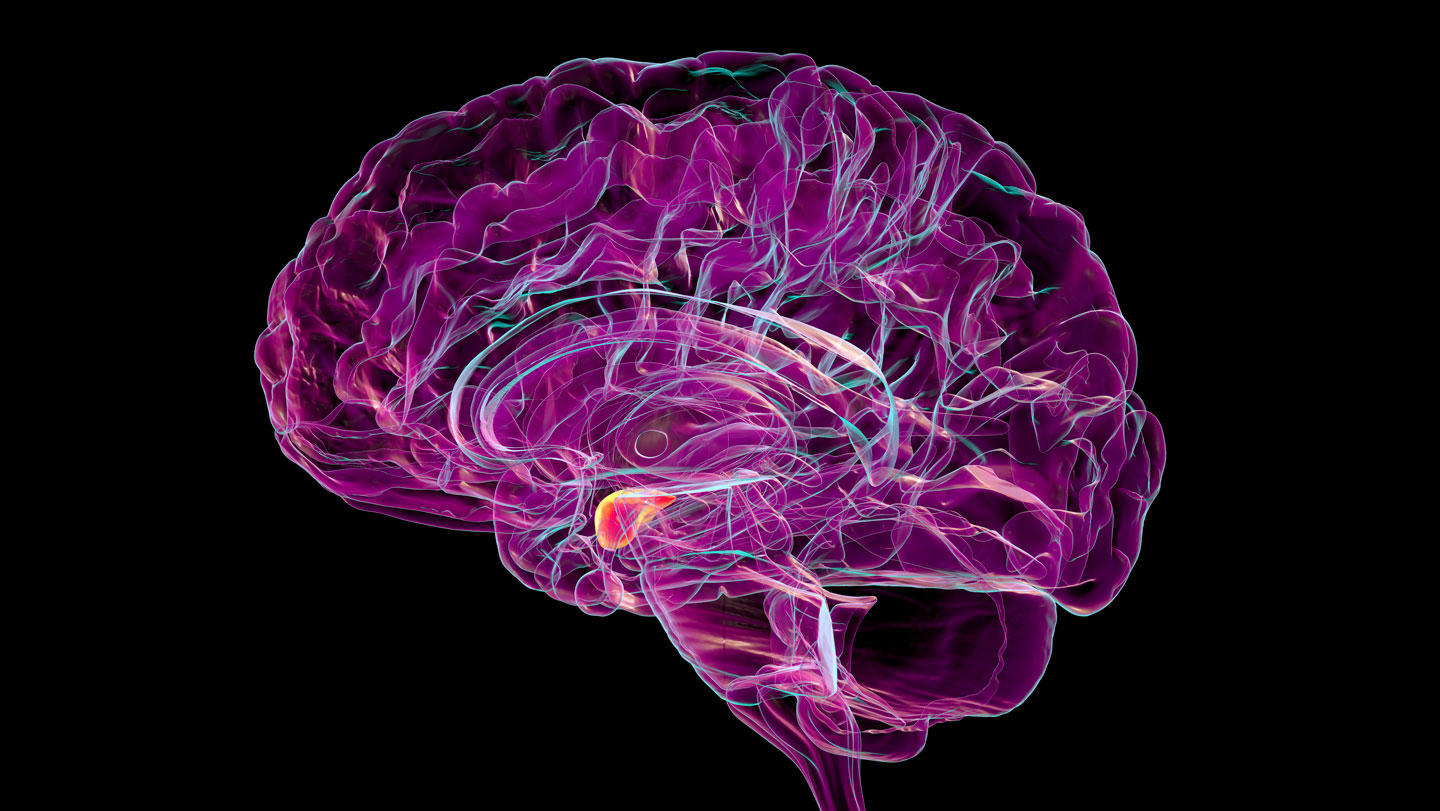
The walnut-sized fear center of the brain also distinguishes the memorable from the mundane.
Neurons in the amygdala act like a save button for memories, scientists report October 27 in Nature Neuroscience. Though the study doesn’t nail down the exact mechanism for memory formation, it points to a broader role for the amygdala within the memory-sorting apparatus.
The study probes an enduring quandary about the mind, says neuroscientist Jiayang Xiao of Cedars-Sinai Health Sciences University in Los Angeles. “We forget much of what we experience … only a fraction of the information we encounter becomes a lasting memory,” she says.
Although a role for the amygdala in memory has been suggested before, previous studies that used electrodes reading brain activity through the scalp, or at the scale of thousands rather than individual neurons, captured a blurrier picture. It’s akin to a microphone recording from outside of a stadium, says cognitive neuroscientist Cory Inman at the University of Utah in Salt Lake City, who was not involved in the study.
“You might be able to tell that there’s a game happening. You might be able to tell when somebody scores a touchdown. You might even be able to tell that one side of the stadium is cheering for one team and another side of the stadium is cheering for another team,” but that’s about it, Inman says. The new study is more like a lapel mic pinned on a single fan in terms of scale and what the researchers were able to capture, adding clarity.
The amygdala, which is typically associated with regulating emotions like fear and aggression, has neurons that are challenging to reach, residing in two clusters deep within the brain. Xiao and her colleagues leveraged electrodes previously surgically implanted in more than 90 people with drug-resistant epilepsy to identify where seizures originate. In their new role, the electrodes singled out neurons involved in saving memories.
Patients were shown images of everyday objects belonging to five categories such as food and animals. About 10 to 30 minutes later, they attempted to recall which images were already shown. Xiao and her colleagues found that activity in certain amygdala neurons corresponded with an image being remembered. The image had been earmarked for longer term storage, Xiao says.
Although participants had to recall something from half an hour ago at most and the exact mechanism for saving memories is still unclear, the researchers think there are two main steps of the process. The team showed that individual neurons have a preference for one of the five categories of images. First the image is categorized by the amygdala. Then, the strength of the category neuron response the first time the image is shown dictates whether the image is later recalled.
A better understanding of this mechanism may pave the way for more effective treatments of memory disorders, Xiao says, a medical conundrum for which there are currently no broadly effective treatments.
The hippocampus is typically more directly linked to memory, so “it surprised me that we observe such a clear relationship between memory formation and activity in the amygdala,” Xiao says. By contrast, the correlation between hippocampus neuron activity and image recall was weaker and slightly delayed.
Future work like observing the impacts of stimulating amygdala neurons on memory will help researchers discern if this region can be targeted to improve memory, Xiao says.
For now, the secret to memory’s hidden machinations remains in the shadows.
Read the full article here